Kidney cancer patients are stunned by their diagnosis, anxious to make a treatment decision, and simply not know what to expect. If you are struggling with the issue of surgery to remove the tumor/kidney or to start with a med, you need to read this. Deb Maskens, Kidney Cancer Patient and Patient Advocate, our guest writer isa valued member of our disease community and currently serves on the Renal Task Force for the National Cancer Institute. A series of links below will also be helpful. (My extra comments will be in italics, like this. )Welcome aboard, Deb!
Clinical Trial Opportunity for Newly Diagnosed (Non Metastatic) Kidney Cancer
As a community of kidney cancer patients, we hear from newly diagnosed patients looking for treatment options. This is written for those patients, and for patient advocates who help patients navigate through their treatment decisions.
The challenge: this clinical trial is available at many locations across the U.S. and Canada, but patients must ask about it BEFORE they have a nephrectomy. Their own doctors may be unaware of the trial and how to work with the trial centres. In many places, patients get booked for surgery prior to learning about this option. That would be too late for a trial like this–it gives a drug therapy before the surgery for a brief period. (In one of the t wo arms, there is medication before the surgery.)
Why Might Patients Consider this Trial?
For years, the standard of care for early stage kidney cancer has been to remove the tumour surgically, sometimes with the entire kidney–either a partial or full nephrectomy. That was the end of treatment and the beginning of surveillance to watch for any signs of recurrence. (And early stage tumors can be quite large–up to 7cm or about 2 3/4″.)
Now we hope to prevent a recurrence of disease. Since advanced or metastatic kidney cancer is still incurable for the vast majority of patients, this is a worthy goal. With preventive or ‘adjuvant’ treatments, maybe we can stop the disease before it gets to the lungs, liver, bones — to those places where it begins to threaten our lives. Other cancers use this approach and offer patients a real chance to avoid recurrence.
Adjuvant – and Perhaps One Step Better to Neo-Adjuvant
We’ve seen trials for “Adjuvant” (or preventative) therapy which hope to prevent recurrence (treatments given immediately after nephrectomy). But one trial goes one step better – it’s for “Neo-Adjuvant” (before nephrectomy) as well as Adjuvant (after).
Patients may want to rush to surgery to “get it out”. In reality, those tumours have generally been growing slowly, undetected for many years. Kidney cancer surgery is rarely an emergency. There is usually time for a second opinion and to check out any newer approaches.
Here’s the thought: given that the tumour cells have gone undetected and tolerated by the immune system for so long, can put those millions of cells to work and make them “show their calling cards” to our immune system before we take them out?
Combining Neo-Adjuvant and Adjuvant Treatment – PROSPER-RCC
The Phase 3 clinical trial called PROSPER-RCC (NCT03055013) is for patients whose tumors are 7cm (2 ¾”) and larger in size, but not spread beyond the kidney area. These patients are at greater risk of spread of the cancer than those with Stage I or with smaller tumors.
Based on earlier studies, nivolumab (Optivo) is now approved for advanced kidney cancer. This is a trial to test whether there is a benefit when nivolumab is given immediately before and after a nephrectomy when tumor cells might have spread outside the kidney but are too small (microscopic) to see on scans. (Typically a patient without spread of disease would not be treated, but monitored.)
The Rationale for PROSPER-RCC: Why It Might Be Helpful
Here’s what I’ve learned:
Checkpoint inhibitor treatments with PD-1 blocking drugs like nivolumab seem to work best when the immune system may be being turned off by this cellular growth pathway. Cancer is deceptively clever and some tumours can express a protein, PD-L1. This protein can turn off our immune cell responses that recognize and fight the cancer. There was a hint of this with some positive data that indicates that these drugs work best in patients whose tumors were “PD-L1 positive”. (PD means Programmed Death and PD-L Programmed Death Ligand or connector. Death to the cells, and the signalling loop that hinders the immune response.)
In theory, when the kidney tumour is in place, there are millions of cancer cells. All of those tumour cells send off multiple negative signals to the immune system to stop it from working. However, if a checkpoint inhibitor was used and stopped those blocking signals, the immune system would have a big wake-up call – e.g., lots of targets with which to build an army of T cells. In theory, these newly educated T cells would later turn into memory cells. (If the body can maintain these memory cells, they would continue to fight any return of disease.)This is much like what happens when we are exposed to certain bacteria or viruses. Once we get exposed to the bug, we don’t usually get it again. Our immune cells have learned (“immunity”) how to kill it more quickly the next time before it turns into a full blown cold. Similarly, if these anti-RCC immune cells ever see one of these tumor cells anywhere in our bodies again, they would know to attack and kill them even if there is no drug in the patient and has not been for some time.
Surgery is still the main treatment to control early stage kidney cancer. But it will also remove the majority of targets (PD-L1) that the checkpoint drug uses to rev up the immune system. Giving the checkpoint inhibitor before surgery may maximize/optimize the drug’s ability to wake up the immune system and build that T cell army.
So the surgery is important. But let’s assume a few cells might be still circulating and have gone undetected for some time. They could still show up later on a scan as an enlarged lymph node or spot somewhere. A boost of the same checkpoint inhibitor right after the surgery could then be used to remind the immune system to continue to look for those cells and kill/eliminate them when they are small. In theory, the immune system will remember what the past “trouble” was: “Hey, haven’t I seen you before?”
From what I understand, this theory worked well in mice. The checkpoint inhibitors worked better if the primary tumour was there to help provide “a target” to activate the immune system first before the tumor was removed. While we’re not mice, this makes sense, no?
Trial Design: What Really Happens to the Patient in the Trial
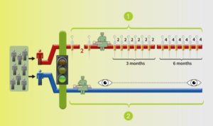
PROSPER-RCC will place patients randomly into two groups:
Group One gets two infusions of nivolumab before surgery (at about 28 days and 14 days before surgery). Following that nephrectomy, the patient will receive more infusions of nivolumab. This is for 9 months post-surgery altogether, with 12 more doses.
Group Two gets the usual standard of care: upfront nephrectomy, partial or radical nephrectomy, and will be followed by close observation at an expert centre.
Two arms/groups: BLUE arm with surgery and monitoring by the trial team, the standard of care; the RED arm with medication before to surgery, followed by more after the surgery.
It is important to note that no patient on this trial receives any intravenous placebo/inactive treatment. Every patient is treated. Each patient will have either the experimental treatment or the standard of care. All are under close observation at the trial centre. This trial has been designed and discussed with patient advocates and is supported by the NCI.
or call the office of the Principal Investigator, Dr. Lauren Harshman, at: 617-632-2429
Deb’s Disclaimer:
As a patient and advocate for kidney cancer patients, I have been delving into the world of clinical trials and trying to understand as much as I can. I’m not a scientist, but I am a patient with this disease, so I bring that lens, along with some abilities to translate science into understandable terms. As a volunteer, I have no financial interest in this trial or any specific medications. @DebMaskensKCC; dmaskens@rogers.com
With the headline, “Nivolumab Shows a Substantial Objective Response Rate in Refractory Non-Clear-Cell Renal Cell Carcinoma”, the article should be welcome to all of those in the in the non clear cell RCC world. Clear cell is the most common, the garden variety of renal cell carcinoma. This is welcome news, as the non clear cell patients get very little attention from the research world. Though the patient with nccRCC might interpret this as, “Good news! Now that they know what to do for me!” , it is just not the case. Rarely is the news all that good or all that simple.
Let’s back up here and lay the groundwork. Clear cellRCC, or ccRCC is the most common of about 10 RCCs. They all land in the kidney, but can vary widely. ccRCC may be about 65% to 85% of the cases of kidney cancer, with the rarer non-ccRCCs making up the rest. Maybe 15-35% of the RCCs are considered rare, with the most common Papillary Type I, Papillary Type II, chromophobe, clear cell papillary, collecting duct/Bellini’s, medullary, translocational (not to be confused with transitional, etc, etc.) and to make it still more confusing, unclassified RCC. But when the most common is described as either 65% of the whole or 85% percent, you have to question if there is clarity in that category!
Clinical trials for RCC have usually only included patients who had clear cell. The reasons are simple; it is the biggest group, the patients can be more readily found, and that is the largest group in need of the medications. But the patients with nccRCC are really also terribly underserved. Back in the day, none of us had many options beyond surgery, so little distinction was made. The prognosis was grim all around, once the cancer had spread.
But the new world of precision medicine, in its name alone, reminds us that the meds need to be developed more precisely, that they be given to the right patients at the right time. The general crap shoot or “wild-ass guessing”, as a friend says, still remains. The latest (not necessarily greatest) group of meds are the newish immune therapies. You have seen their ads, no doubt.
One of those is Opdivo or nivolumab, its research name. It tries to unblock some of the inhibiting mechanisms that prevent the immune system from doing its job, but it has been tested in trials only with clear cell patients. BUT, that does not mean that only clear cell patients are being prescribed the meds–this, thanks to the slightly wild west of the US medical system, that can truly go beyond the FDA approved medication guidelines.
This study, which will be formally presented at ASCO in June, 2017 was announced with the headline above, “Nivolumab Shows a Substantial Objective Response Rate in Refractory Non-Clear-Cell Renal Cell Carcinoma”. The researchers are NOT in charge of the headlines, so we must dig deeper and see what this study really means to the patients with nccRCC
I tried to sort out what it means–or does not mean. My quick review is that it does not give a great deal of clarity to the majority of those nccRCC patients. A more complete report may improve upon this. Based on this link, I offer the following:
“I am always concerned that these new study reports are characterized carefully. They are always more complex and incomplete than I would like. A patient in a forum says this tells of ‘good’ responses, and especially so for the non clear cell group, but s does ‘good’ really mean generally a benefit to those rarer nccRCCs? Until a fuller report emerges, I can only note the following:
There were 23 patients, from three centers, with a median age of 59. Surprisingly 30% were African -American. This may tell us that there are more African-Americans with the rarer non-clear cell RCCs, or could reflect the local population of the three centers. Only 23 patients and with a mix of diseases will never meet the statistically critical requirements to reach the level of excellent evidence–but it may be all we have at this point.
All 23 had non-clear cell, but nearly half had ‘unclassified’ RCC, quite a high rate. Usually that is considered to represent between 1 to 4% of renal tumors. Most of the rest were papillary, but they generally make up the largest percentage of nccRCCs. No distinction is made here between Papillary Type I or Type II, which are really quite different diseases. Papillary Type I and II are the most common of the uncommon, non clear cell RCCs, and are readily distinguished from each other. This would be valuable info, and wonder if this was noted in the fuller report.
Only 3 of 4 patients had nephrectomies before the trial treatment. Were 1 of 4 patients too sick to be given the standard of care of surgery or were their doctors unaware of that? How does this affect the study, and were the no nephrectomy patients from one center or with one subtype? We do not know the reason for this high rate of no surgery, and at a time in which it is clear that the removal of the tumor is a great benefit to the patient, metastatic or not.
Two-thirds had metastatic disease at the time of diagnosis. Of the total 23, 74% had a prior treatment, mostly Sutent or Votrient. Of these patients with prior treatments, 26% had TWO such treatments. Thus these patients had already received treatments that were not directly approved for their subtypes. This is not too rare in the US, where we have greater leeway from our prescribing doctor than do patients elsewhere. But how does this fit in with the relatively low rate of nephrectomies?
This report does not say how quickly they were treated, i.e., how long from initial diagnosis until treatment with Nivolumab?A patient with Papillary Type II found to have no metastatic disease at the time of diagnosis, but who received a nephrectomy, was monitored for a year or so, then went on one or more systemic therapy is quite different from the patient with an unclassified RCC, metastatic at the time of diagnosis, not given a nephrectomy, though treated quickly with Nivolumab. What can be learned when there are such wide variations in just 23 patients that would be helpful to the Papillary Type 1 patient?
The follow up period was a median of 6.5 months, which seems very short, especially when the median Progression Free Survival of the responders was 4.2 months. The median OS is not given. That certainly may reflect an ongoing study situation, or a failure to provide a longer period of follow up.
As to objective response, 6 of the 21 evaluable patients (29%) had a Partial Response, which would likely be a 30% reduction in metastases. Another 4(19%) had Stable Disease. Two of the 23 patients died, but not from the treatment. (Assume that had to be due to the disease, but certainly indicates that for nearly 10% of the patients, this was not at all effective.)
When the final analysis was done, nine patients were still receiving Nivo. Newly recruited patients might still be in treatment at that time, but those recruited earlier may have gone out of the trial at the same time. It is important to not that Nivolumab treatments were stopped in three patients due to intolerance, and six more had postponed treatment, i.e., 9 of the 23.
Certainly we need to find meds which create responses for nccRCC patients. However, I am concerned we draw any certain conclusions from this study. Indeed, it is “good” to know that the treatment was tolerable for the majority of the participants, but not so good to read that 6 of the 21 patients had to postpone treatment, and three were removed from treatment due to intolerable/toxic side effects. We also do not know which subtypes seem to have shown responses, which would have been qutie easy to report. Did the group with Papillary Type II do generally better that the majority “unclassified” group? No answer from this stury report. And in the back of my head, I keep wondering why in the world there were so many unclassified patients in this small study? Was there a standard pathology review, or could these patients been misdiagnosed by one pathologist. Typically there is a single pathologist which can standardize the reporting. Were all these patients properly diagnosed?
Just wishing there were greater clarity and hoping to get a fuller report, post ASCO.
Without a doubt, the ‘good’ that comes from this sort of report begins with the recognition that the nccRCC group is underserved by the research community> They probably have the poorest outcomes, rarely have a clear diagnosis, and must wait for the ever popular “further research is warranted.” But all must be aware that these very small observational studies must be reviewed very carefully for what they show or do not show. Again, one to watch at ASCO, but not enough to make a major change in treatment for any one with a non clear cell RCC.
PS. Does your doctor know that there are at least four subtypes of clear cell–the big ‘common’ group–which have clearly different survival patterns? Thought so.
Recent headlines called a new medication, Nivolumab, both a miracle or breakthrough and more. Is it hype or hope?Why is it so hard to sort out the reality?
Let’s go through the facts from the New England Journal of Medicine and ignore the headlines. First, its being in the NEJM is important, as it has passed review by other researchers. (Sadly missing in too many ‘breakthroughs”).
The new med, Nivolumab was compared against Everolimus, a second-line treatment. Therefore Evero is thought to be of lesser effectiveness than the first line meds. Second-line meds are generally used when others meds quit helping or their side effects are too hard. Automatically NOT the miracle cure, but another option when first-line treatments fail.
Should Nivo have been compared to the first-line meds? Being better in the first-line would be bigger deal, but we need more approved meds. Second-line treatments usually are easier to ‘beat’, as the new med must be better or less toxic. Again, more likely to be approved!
PATIENT CHARACTERISTICS
The study had 821 patients 24 countries, half using Nivo and half Evero. Patients were similar, 90% having had a nephrectomy, removing the tumor and some or all the kidney. Then the cancer spread, making metastases, (mets, for short). These patients had 1-3 treatments, first-line drugs like Sutent, a targeted therapy, and a few had used cytokines or even chemotherapy. Having had an mTOR inhibitor like Everolimus was not acceptable. Most had lung mets (67%), followed by liver(12%) and then bone mets (18%). Most with 2 or more sites of mets.
To enter the trial, the patient had to have had disease progression after their last treatment, within six months of enrolling in the trial. No doubt, some patients had greater disease progression than others, but had relatively good performance status, not completely bed-ridden or unable to function.
The median time from initial diagnosis of kidney cancer at any stage to entering the trial was 31 months; half had been diagnosed less than 31 months ago, and half more than 31 months before the trial. That range of time from diagnosis to trial was 1 to 392 months. That means that for some patients, they went a long time either fighting the disease since diagnosis, having a later recurrence, being treated, yet having disease progression years after the intial diagnosis. At least one person was diagnosed 392 months earlier. This is a good reminder to patients who have been told, “I got it all”. This darn stuff can return, so having a plan B is important. Again, the previous treatment failed and these patients got directed into this trial.
GENERAL RESULTS
Median Overall Survival (OS) is a measured when one-half of the total number in the group dies. Median OS for Nivo was 25 months with some patients still surviving at time of report, beyond the 25 months. For Evero, OS was 19.6 months, some of who were also likely surviving, as well. The OS of 25 months was clearly better with the Nivo group by this analysis. Nevertheless, half of all the 821 patients total died while on this trial from progressive disease. Of 183 of the 410 Nivo patients, 183 has died by 25 months, and 215 of the 411 Evero patients had died at 19.6 months.
There is no report of ongoing response here, but many went on to other meds, as explained below.
Median Progression Free Survival (PFS), measurable growth of disease, was 4.6 months for Nivo, 4.4 months for Evero. The median shows that half of each group, roughly 200 each had return of disease in less than 5 months! Again, these trial patients were pretty sick or at risk. All had been treated earlier, and had to stop previous treatments due to recurrence of disease. However, this shows a pretty quick return of disease or new growth from the base CT scan for nearly 65-70% of all patients.
One subgroup did a bit better than the 4 1/2 months median PFS. At six months after the start of treatment, there was a special subgroup was noted, about 1/3 of those patients–145 pts (35%) with Nivo, and 129 (31%) with Evero. Obviously they did not die or have Progressive Disease until after six months. The Nivo group had eventually had a median PFS of 15.6months, and the Evero group, 11.7 months. Their success pushed the median OS higher, especially for the Nivo group.
Obviously, there were some patients with far more aggressive disease in both groups, some dying before six months, and others not progressing to more disease until after six months. In contrast, nearly 1/3 of all the patients had PFS of 12-15 months, and much longer OS. What is the common characteristic in the most successful two groups in both arms of treatment? Not answered by this trial report.
The duration of treatment was longer with Nivo, and likely easier to tolerate. Since Nivo was given by IV every two weeks, the doses were most consistently received. Even so, 51% of them had dose delays, but no per dose reductions. Those people were seen by the medical team every two weeks.
The Evero group took oral meds, and 66% had dose delays or interruptions with 26% with at least one dose reduction. This would indicate that these meds could be hard to take, or perhaps lacking the same interaction with their medical team. Of course the Evero patients may have underreported how much of the medication they actually took!
However, the reported types of side effects were generally similar, but the more severe grade 3 and 4s effects in the Evero group.There were 2 treatment related deaths in the Ever group, none in the Nivo group.
POST PROGRESSIVE DISEASE
Even after the disease did progress, about half of patients in both groups stayed with their meds–despite ‘failing’, the researchers hopes that would continue to benefit, perhaps slowing the disease. In a local clinic setting or with a less experienced docs, their meds might have been stopped or changed. Afterall, those meds were no longer “working” and mets are growing. This approach is significant to consider, especially after multiple treatments. (The decision to keep giving a medication or increasing its dosage where tolerable is causing some changes in treatment in a number of the targeted therapies.)
Perhaps because of being in a trial or getting care than was more expert than most, one-half of patients chose to keep on the trial meds. Others crossed over to the med in the other arm or returned to existing non-trial meds. In some countries, there were likely fewer choices than in the US. There are no real stats as to survival for those on those who stopped taking the meds. It is reported that indicate that 55% of the surviving Nivo group and surviving 63% of the Evero group went on to other agents. About one-quarter of the Nivo group shifted to the Evero. Of the Nivo group, 36% shifted to axitinib.
Sadly, as per the chart in the New England Journal of Medicine, all these patients had died by 30-33 months post enrollment. However, it is again not clear what was effect, if any on that period from the non-trial drugs. Of the 227 who stopped Nivo for any reason, nearly half shifted to Evero. Of those who stopped Evero, 140 went to Axitinib.
DURABLE RESPONSES? HOW LONG? FOR HOW MANY?
The writers of the study say that there was a higher number of objective responses with Nivo vs Everolimus, and that many (of the Nivo group) “were durable”. There is no definition of ‘durable’. My question is “What equals durable?”. We patients really want a cure, but are very grateful for anything that pushes the cancer back, slows it, stops in from growing any further. Nevertheless, we do want those responses to last. The clearest reference to durable responses is a note that 32 of the Nivo patients and 6 of the Evero patients had a response that lasted more than 12 months. But in an unexplained statement, the median duration of treatment was just 5.5 months for the Nivo patients, 3.7 for the Evero group. It seems that there was not an extension available, or that the patients moved on to a different treatment or passed away.
CONCLUSIONS AND EDITORIALIZING AGAIN
It seems that Nivo is more helpful for some patients than others in this group previously been treated with other TKIs. This is NOT A SILVER BULLET. There would be greater value to know more about the molecular nature of the tumors of the responding and the non-responding patients. We desperately need to know for whom any of these drugs is likely to be more effective. The headlines that don’t discuss that challenge underserve us, as does the design of the trial that does not elicit the more nuanced, genomic data that could be forthcoming!
We all know that headline claims are more wonderful than the reality. The story of RCC medication development is that of more and more help in a difficult disease, making mixed progress, while the other researchers find out that RCC is really many diseases. Clear cell is probably better defined as being made up of four types, Papillary Type 1 and Type 2 being further divided into three Type 2, then there is chromophobe, clear cell papillary and the really odd versions of RCC. I known this, and so do you. But why don’t the researchers incorporate those definitions and monitor the patients with those various subtypes as they go forward?
Is there another way to measure the benefit from any medication?
We all want the cure, the Complete Response (CR) that can lasts many months or years. Often we have to settle for some reduction in our tumors or mets, a Partial Response (PR). But even “Stable Disease” is welcome news. To get that cancer back in its cage, even for a time, is better than “Progressive Disease”. When the cancer is progressing, your life may be regressing, and that isn’t what you want to hear. That Progression Free Survival (PFS) has to start with stopping the cancer.
As complete and durable (ten years) responder to high dose interleukin 2 (HD IL-2), I welcome any discussions of “Clinical Benefit (CB)”. CB includes all the good responses with any cancer treatment, CRs, PRs, and SDs. We and our doctors need this information to make informed decisions about treatment, for IL2 or other meds. The value of Stable Disease has been ignored in many studies. Maybe there are lessons here for you and your doctors, especially about the under-utilized HD IL2.
Clinical benefit (CB) of high-dose interleukin-2 (HD IL-2) in clear cell (cc) metastatic renal cell carcinoma (mRCC).
There are few new studies about the use of HD IL2 following the approval of the targeted therapies. The ease of use of these agents, along with the desire not to send patients to specialty centers for IL2, limited its use. It was difficult to select patients, and the CR and PRs were relatively small in number. Doctors often did not discuss the possibility of a cure with their patients. Did patients also miss the chance for Stable Disease, and with it, a “Clinical Benefit”?
Patients in this study who did not have a CR, but whose cancer stopped growing benefited. That CB was not counted in terms of the approval of the drug, nor do doctors consider it in their recommendations. Should this possibility be discussed with patients? Most patients would surely answer, “Yes!” to that question.
The researchers recognize of the value of Stable Disease (SD) as an outcome, versus only Complete Response (CR) or Partial Response (PR). The usual outcome measures, Progressive Free Survival (PFS), or Overall Survival (OS), are noted, as isTime to Next Treatment (TNT). TNT implicitly recognizes that a failed or limited response will likely be followed by another treatment. Early on, there were no subsequent treatments, sad to say.
The original clinical trial which led to FDA approval of HD IL2 recognized only CR, which was 5%, with the median not reached during the trial, and PR, which was 14%. Study footnotes indicate that three of the PRs had surgery which rendered them disease free at the time of the publication. This would now be called a “salvage therapy”, and put them in the No Evidence of Disease (NED) class. A different analysis of this data would have upped the CRs some small percentage, and some SD would also have been found.
Also the definition of PR was 50% or greater reduction in measurable tumor size, the sum of the perpendicular diameters of all lesions, with no new increase of sizeof any other mets. Far less strict measurements of PR were used in the targeted therapy trials, with a 30% tumor reduction defined as a Partial Response.
With those definitions in mind, note that there are CRs in 11% of patients, with a PR in an additional 6% of patients. Most important is the SD category, which was achieved for 31% of all patients. This total of 47% is described for the group as being of Clinical Benefit (CB). Certainly patients value the responses of SD, which seems to have provided slightly over one year versus 3-4 months benefit to those who did not have SD.
When comparing the value of Objective Response (OR) with its median of 1616 days to that of Stable Disease (SD) measured as 1476 days, one can clearly see the value of achieving Stable Disease. Unfortunately, those patients with Progressive Disease, or with responses Not Evaluable (NE), showed OS of 365 days.
Patients should be aware of these definitions and the impact the lack of parallel comparisons in making these critical decisions. Ten years ago, the patients reminded one another to stay alive until the next treatment. Having Stable Disease made that possible. Let’s apply the same tests to all the available treatments when making these life-changing choices of treatment.
Author(s): Neeraj Agarwal, David D. Stenehjem et al University of Utah, Huntsman Cancer Institute, Salt Lake City, UT; Comprehensive Cancer Centers of Nevada, Las Vegas, NV; Pharmacotherapy Outcomes Research Center, College of Pharmacy, University of Utah, Salt Lake City, UT
Background: HD IL-2, an immunotherapy, is a standard of care for a select group of patients (pts) with mRCC. Generally objective response (OR) rates, i.e. complete response (CR) + partial response (PR), of 16-20% are discussed with pts, but not disease stabilization (SD). Recent data suggest that cancer immunotherapy may improve survival without inducing OR. Thus, treatment with HD IL-2 may provide survival benefit to an additional group of pts not experiencing OR, but only SD as the best response. Here we report CB (OR+SD), and specifically report outcomes of cc mRCC pts experiencing SD as the best response, on treatment with HD IL-2.
Methods: All sequential cc mRCC pts treated with HD IL-2 at the University of Utah Huntsman Cancer Institute from 2000-2012 were included. Pts were evaluated for best response, progression-free survival (PFS), time to next treatment (TNT) and overall survival (OS). Two practitioners independently reviewed HD IL-2 response with discrepancies adjudicated by a third reviewer.
Results: 85 pts, 79% male, were identified with a median age of 56 (range 32-76) years. Pts belonged to the following MSKCC risk categories: 11 (13%) good, 70 (82%) intermediate, and 4 (5%) poor risk. A CR was identified in 9 (11%), PR in 5 (6%), SD in 26 (31%), progressive disease (PD) in 38 (45%), and unknown/not evaluable (NE) in 7 (8%) pts; yielding a clinical benefit in 40 (47%) pts. The median PFS, TNT, and OS in these individual groups of pts are compared in the table.
Conclusions: A clinical benefit of HD IL-2 was achieved in nearly half of all clear cell mRCC patients. OS was not significantly different in OR and SD groups. Even though OR favorably determine outcomes, SD is also an important response criterion, and may be discussed during counseling patients for treatment with HD IL-2.
PFS, days
TNT, days
OS, days
Overall
152
264
817
SD vs PD and NE
337 vs 78 (p<.0001)
373 vs 110 (p=.0001)
1,476 vs 365 (p=.0003)
CB vs PD and NE
791 vs 78 (p<.0001)
735 vs 110 (p<.0001)
1,616 vs 365 (p<.0001)
OR vs SD, PD and NE
NA vs 99 (p=.0003)
953 vs 166 (p<.0001)
1,616 vs 603 (p=.0021)
OR vs SD
NA vs 337 (p=.0234)
953 vs 373 (p=.0015)
1,616 vs 1,476 (p=.2094)
Abbreviation:PFS, Progression Free Survival; TNT, Time to Next Treatment, OS, Overall Survival; NA, not achieved;SD, Stable Disease; PD, Progressive Disease; NE, Not Evaluable; CB, Clinical Benefit;CR, Complete Response; PR, Partial Response;OR, Objective Response
When you are suddenly thrust into the medical world, unwillingly and without any kind of road map, you are surrounded by poorly marked turns, meaningless abbreviations and the sudden shift in the dialect. The Wellness Center is usually about having lost one’s “wellness”, a word used only in the medical world, and not by real people.
Pressed to make decisions that may change your life, for the better or worse, you can be confused by those clever new words, some from the marketing people (see above) and others from the clinical side. It is critical to understand how familiar words get reworked to explain new concepts. Such explanations rarely reach patients, who are numbed and deafened after a shocking diagnosis. And in the medical “new-speak”, those same patients may be told that this is the time in which they must take charge of their health, and make wise decisions quickly and correctly. I find this a cynical and self-serving approach, as rarely is any real education offered in the language of the patient.
In kidney cancer, we have been blessed with new drugs these past eight years, but have no clear way to determine which of these agents might be of benefit to any of us. On top of the shock of diagnosis, the patient is thrust into a guessing game. Even the doctor is forced to play along, and often neither party knows the rules or the chances to win. The doctor may recognize the vocabulary used in this new guessing game, but the patient does not. Words which have meaning in day to day life don’t work the same. Even some of the goals of the game are unclear to the patient. Wait! You probably think that being cured is the goal. you
For example, we patients think that “progress” is good, but that is not true in cancer. Progression is the goal of the cancer, so Progression Free Survival (PFS) measures the time between treatment and when the cancer is on the visible move again. The word “visible” is important here, as that is a reminder that cancer does not just start at a size or style to match the sensitivity of imaging. X Rays cannot see things as small as a CT scan can. Bone scans see bone mets better than other scans and so on.
In reading clinical trials, you will encounter “durable” to explain how long a median PFS can be. It may be described as remarkably durable, but in the pre-patient world, we would think that is pushing into years and years. In reality is may be 15- 18 months. We happily grasp at any more months than the non-treatment reality may be, but be aware of your and your doctor’s expectations in durability.
“Durable response” is surely what we want, but that is not translated to a cure, which might be the patient’s interpretation. When you hear that, do ask for clarification, “How long does that response last? What do you mean by ‘durable’? What do we do after the duration of response comes to a stop?”
Having a firm grasp of this term and all others is an absolute necessity, and even if that is hard–in the real sense–it will be worth it to you. You will have greater understanding of the treatments, the disease process, and a bit more sense of where you are.
More on these topics later, but do track the language, and remember than you still speak the old language. At the very least, be ready to question anything that has that new dialect sound to it!
I was dying ten years ago. My kidney cancer had moved into my lungs, threatening to choke me to death.The tumor and kidney were gone, but 100s of tiny lung metastases were growing. Lucky to get an FDA-approved immune therapy, high dose interleukin 2, my own immune system was revved up so as to destroy the cancer. Thus, I am intrigued by all things about the immune system and cancer research. “Adaptive immunity in cancer immunology and therapeutics”is one of the most comprehensive explanation of the tumor cell/immune system interactions–that I can somewhat(!) understand.
My summary is below, a more patient-friendly version. Don’t hesitate to take on the original, via the link! It is just the kind of article to take to your doctor to discuss immune response meds/treatments. It begins with the “abstract”, a summary of the information to follow.
Abstract: The vast genetic alterations characteristic of tumours produce a number of tumour antigens that enable the immune system to differentiate tumour cells from normal cells. Counter to this, tumour cells have developed mechanisms by which to evade host immunity in their constant quest for growth and survival. Tumour-associated antigens (TAAs) are one of the fundamental triggers of the immune response. They are important because they activate, via major histocompatibility complex (MHC), the T cell response, an important line of defense against tumourigenesis. However, the persistence of tumours despite host immunity implies that tumour cells develop immune avoidance. An example of this is the up-regulation of inhibitory immunemonoclonal antibodies in clinical practice have been developed to target tumour-specific antigens. More recently there has been research in the down-regulation of immune checkpoint proteins as a way of increasing anti-tumour immunity.”
Immune Responses in Tumors—A Quick Summary by Peg
Since cancer cells are genetically different from normal cells, they also produce different substances—antigens—which can make them more noticeable to the immune system. Any antigen will generate a response from the immune system—think how the body reacts to an infection, an insect sting or a splinter.
Antigens trigger the immune system into action, keeping abnormal cells from taking over the system—most of the time. To grow, tumor cells develop inhibitory responses to limit or down-regulate those immune responses. An over-active immune response can be problem, well-known to those with severe allergies or auto-immune diseases like lupus. Keeping the proper balance is the norm for the immune system, despite ongoing external and internal changes
Using knowledge of these interactions to support the immune system, researchers have develop agent/medications. These are intended to strengthen the beneficial responses, and to prevent the tumors from suppressing or down-regulating those desired responses. Some monoclonal antibodies can effectively target these tumor-specific antigens and trigger tumor death or inhibit such growth. Some of these new agents include bevacizumab (Avastin), rituximab (Rituxin), alemtuzumab (Campath or Lemtrada), bortezomib (Velcade), denosumab (Xgeva) and trastuzumab (Herceptin), among many others, and for a variety of cancers.
Be aware that these agents may be called by the brand name, as Sutent, or the scientific name, as sunitinib, and may have several brand names for different cancers. Just another new challenge to all of us newbies.
Tumors exist with a system of structures, various types of cells and with a chemical signaling process. These shifts away from the normal cells and organs produce tumor antigens. The immune system notices the antigens and works to destroy the foreign cells. Then the tumors shift to counter the immune response in an endless signaling battle. It is a dynamic “fail-safe” system, with multiple checks and balances, work-around pathways, evasive signaling, and constant testing to maintain itself. When this system does fail, a tumor can be established and move to different sites.
Solid tumors have a tumor core, a margin that is invading into a healthy structure–blood vessels or layers of an organ–and lymphoid components. This can vary patient to patient, despite the seeming similarity of tumors, and vary from one metastatic tumor site to another. Inside the tumor will be the immune-cell types–macrophages, dendritic cells, natural killer (NK) cells, mast cells, B cell, and T cells. Different immune cells can be found in different parts of the tumor, and the variation and the density of these cells may play a role in clinical response. It may be that this reflects the robust nature of the natural response to the tumor invasion, or reflect that the system is being overwhelmed by the tumor. Others think that the infiltration of immune cells can be utilized the support of the treatments given to the patient.
The linked journal article goes into detail as to the various types of responses, including adaptive immunity, immune editing and immune evasion. In summary, there are numerous approaches to limit tumor growth within the complex system of antigens and immune responses.
As immune cells infiltrate a tumor, that infiltration can be measured. What is the meaning of a higher or lower level of immune cell infiltration? The following paragraph sums up the challenge of using tumor infiltration as a marker of prognosis or treatment response.
It is a commonly held belief that infiltration of immune cells into tumor tissues and direct physical contact between tumor cells and infiltrated immune cells is associated with physical destruction of the tumor cells. That can reduce the tumor burden, and improve prognosis. An increasing number of studies, however, have suggested that aberrant infiltration of immune cells into tumor or normal tissues may promote tumor progression, invasion, and metastasis. Neither the primary reason for these contradictory observations, nor the mechanism for the reported diverse impact of tumor-infiltrating immune cells has been elucidated, making it difficult to judge the clinical implications of infiltration of immune cells within tumor tissues. J Cancer 2013; 4(1):84-95. doi:10.7150/jca.5482
Tumor Infiltrating Immune Cells—a Good Sign or Not?
If the immune system is at work, immune cells infiltrate the tumor to work directly against the tumor cells, is the tumor destroyed? Does the body naturally destroy the tumor? Does the patient benefit from medical treatments which support the immune system? Unfortunately, the presence of the tumor-infiltrating cells can mean very different things, with a better prognosis in one type of cancer, and a poorer prognosis in another.
Monoclonal antibodies can target antigens in blood cancers and solid tumors. In blood cancers, antibodies counter several cluster of differentiation (CD) markers, and in solid tumors, growth factors such as EGFR (epidermal growth factor receptor) or angiogenesis factors, such as vascular endothelial growth factor (VEGF). The mechanisms of action can lead to direct cell death, or simply impede its growth or inhibit checks on the immune response.
Normal cells are naturally programmed to die, but cancer cells do not “follow the program”. When certain proteins on the surface of cells bind with one another, the expected immune response is inhibited. These anti-PD-1 (anti-Programmed Death-1) proteins bind with other proteins, the binders or ligands, PD-L1 and PD-L2. Studies indicated these agents can help the immune system, with some disease stabilization or tumor shrinkage. Recent trials show some response by 20-25% of patients, some of whom had failed previous treatments. Some responses lasted more than a year. In a few cases, some responses were lasted for a period after stopping the medications. Newer trials will likely combine several of these therapies. This is not without risk, as some had severe side effects, and several patients died from such side effects.
Nevertheless, the earlier successes with this approach and the increased knowledge of the various immune responses to be targeted will continue, especially in combination studies. This work will have impact on existing immune therapies, as does the more integrated approach to cancer treatment.
I welcome any comments and corrections, and remind you that I am a patient, and am not a medical professional. My goal is to help educate other patients to receive the best understanding of their illness and best possible treatment.
“Now what?” may be the first coherent question a newly diagnosed cancer patient asks. Maybe the smarter version of that is “What–when and why?” And your doctor had better have a good answer, as to the treatment, the when and the why.
We cancer patients usually get surgery “first”, even when the disease has spread. Primary surgical strike and then a clean-up operation, in the ‘war on cancer’ parlance, we think–when we can think. “But which is the best and first clean-up approach?” we must ask. “What works the best? What can I take with my other health problems? Where does surgery or radiation fit in this scheme? What does the doctor favor and why? Where do I get this treatment? And then what?”
Treatments and their sequence are often chosen with little reliance or clarity as to the data. But some light was shed today at ASCO (American Society of Clinical Oncology). It released a comparison of the sequencing of High Dose Interleukin2 (HD IL2) and of targeted therapies for metastatic RCC. Which should come first?
It shouldn’t be a high-stakes gamble to choose a medication, as no one can guarantee any results–with any of the meds. You take a chance with any drug, so which do you start wi We may be closer to a logical approach in sequencing these drugs. Sequencing of these highly different medications has measurable effect on overall survival (OS)—and to patients’ lives. That sequencing is critical and certainly can extend life, even when treatments fail, as they so often do.
A retrospective study of 97 US patients who received HD IL2, before or after a targeted therapy was just presented at ASCO. These patients were followed for a median duration 37 months–half more than 37 months, half fewer than 37 months. Of that group, 22% had either a partial (14%) or complete (8%) response to HD IL2. (No specifics as to what was a “partial” response, perhaps a 30% shrinkage of the total tumor burden). In addition, another 24% of patients had Stable Disease(SD). Thus, nearly half of these patients benefited from having had HD IL2.
Stable disease is better than progressive disease, as any patient knows, though it was rarely measured in older trials. Though we patients really want a cure, we do want to be around for the next treatment, to have a surgery or ablation to remove the “stable” tumor, or to try another medication.
Of these 97 patients, 82 received HD IL2 before any targeted therapy. Another 15 patients had HD IL2 following a TKI therapy. That timing made an important difference. HD IL2 followed by the TKI, showed a median Overall Survival (OS) of 61.8 months. The OS of those with the TKI before the HD IL2 was 48 months. A median, not an average, so half lived longer, half lived shorter than the quoted medians.
A pre-2006 NCI (National Cancer Institute) series showed a 19 month median survival for HD IL2 alone, and a similar 19 months for the use of targeted therapy alone. Using the two in sequence dramatically improved OS, especially when HD IL2 was first line of treatment. Obviously things have improved, though it can be very difficult to compare older trial data, as so many variables are different–including the type of RCC the patients had as they entered the trials.
Several points can be made from this study. First, no therapy should be examined only as to Complete or Partial Response. Stable Disease also adds to Overall Stability. To stop the tumor from growing, even if for a period of time, is valuable to patients and can prep them for the next anticipated treatment. Sure beats tumor growth!
Second, therapies should be chosen to maximize their impact on the overall survival of the patient. Some patients will naturally be precluded (or delayed) from surgery, or taking one drug due to existing co-morbidities, due to heart disease or liver damage. For those post-op patients, likely to tolerate the side effects of HD IL2, it should be given in a first-line setting.
The most critical variables that impact patients are the recommendations and expectations of the physician. Most patients are not even told about HD IL2 treatment, or it is dismissed casually as “not for you”. Others are told to wait until more mets emerge, with some weird theory that waiting for more trouble is a good thing! Many nephrectomy patients are not monitored post-operatively, despite the risk of mets. This is surely an indicator of the lack of knowledge by urologists. Still others are told that the disease has spread, and that nothing can be done–also untrue.
The rarity of RCC and its variants leaves most physicians unaware of all options in the field, and how to any one might suit for a particular patient. Most oncologists to whom patients are referred, have little or no experience treatmenting for RCC, or may not access to academic centers for support until it is too late. Even the pathology of the primary tumor and later metastases may be questionable, adding to the challenge of care.
With the dramatic changes in the RCC field, this is to be expected—but not tolerated. The patient may have to provide his physician with the data that can extend or save his life, which is a sad but realistic commentary on the field today.
Most people are not surprised that there is no ONE thing called cancer. Tumors in all the organs or invasive cells in the blood or bones are referred to as cancer, but start when cells go wrong, whatever the cause. As soon as you are told you cancer, whatever it, the quest begins to find out exactly which cancer it is. With kidney cancer, or its more melodious name, renal cell carcinoma, there seem to be endless variations on what may be called kidney or renal cancer. To treat it requires a very careful analysis of what is really is, starting with the pathology of the tumor when it is biopsied. With kidney cancer that biopsy is usually done after surgery for the tumor. That biopsy will describe the shapes and type of cell in the tumor, which can be mix of types. And then the real work begins.
A recent article in “European Urology” reviewed the mix of HEREDITARY renal cancers, those that arise due to one’s background. More common are the “sporadic” kidney cancer that could arise out of the blue or in response to some environmental toxin. There are ten Heredity Renal Cancers, or HRCs. My goal is to alert the reader to the possibility that his cancer might be one of these. This would affect treatment, and may suggest the need to test family members.
If you have kidney cancer or RCC, you may be familiar with “clear cell” or “papillary” to refine the description of the cells in the tumors. This may not be the whole story, as those HRCs—the hereditary kinds—may manifest a mix of ways, including as clear cell or papillary histology.
The most common HRC is Von Hippel-Lindau (VHL) disease, with both benign or malignant tumors. RCC can be found in a 24-34% of VHL patients, appearing at mean age 39 years (far younger than non-heredity RCC), and often with multiple tumors and in both kidneys. Cysts which appear not to be malignant must be watched–they have the potential to become malignant over time. Generally they are managed based on the size of the largest of these lesions. Clear cell RCC is the one VHL-related subtype.
Hereditary papillary renal carcinoma (HPRC) is rarer, and typically occurs later in life. Papillary tumors are the only phenotype with HPRC, and patients often develop numerous tiny tumors, 1000 or more. These tumors are considered type 1 papillary renal cell carcinoma (pRCC) with a low nuclear grade, monitored with CT scans, and some do metastasize, though this is rare. The MET gene is implicated in the growth of these tumors.
Hereditary leiomyomatosis and renal cell cancer (HLRCC) is newly identified as a HRC. Rarely do patients develop RCC, but are susceptible to developing multiple leiomylomas, which are generally benign. When there is early onset of HLRCC, then RCC is found in about 20% of those patients. These tumors can be aggressive, and about 2/3 display a papillary pattern. Such tumors tend to be hyper-vascular.
Birt-Hogg-Dube (BHD) syndrome is quite rare, about 1 in 200,000 people, and thereby likely under diagnosed. This raises the risk of developing kidney tumors, which occurs in 25-35% of BHD patients, and at mean age of 50. These tumors have varying histologic subtypes, generally chromophobe RCC or hybrid variants. And there can be variants in the same tumor or within the kidney. There is a risk of metastases, though rare. The characteristic skin lesions of BHD syndrome are not malignant.
Even more rare is Tuberous Sclerosis Complex (TSC), which can manifest itself in renal lesions, cysts and occasionally, RCC, the latter at a young, average age 28. Neurologic complications can accompany this syndrome.
SDHB-associated paraganglioma/phaeochromoytoma is another heredity condition which may give rise to a mix of renal tumor, including clear cell RCC, chromophobe RCC and oncocytomas, i.e., a mix of histologically different types.
An HRCmay be suspected in patients with a family or individual history of renal tumors, in the instance of both kidney having tumors, or one kidney having multiple tumors or in early-onset renal tumor, i.e., under 50 years of age.
Clinical diagnosis can be further refined by genetic testing, and thorough review by an experienced uropathologist is fundamental to the diagnosis. First consideration would be a VHL analysis and genetic analysis of SDHB and FLCN genes, as warranted. Patients with type 1 papillaryRCC should be considered for MET analysis. The presence of clinical symptoms related to any of the syndromes will guide the gene screening. Testing on family members may well be warranted.
With these cancers, it is possible to have multiple lesions and affect both kidneys. Thus, treatment should preserve renal function and control the risk for metastases. Use of ablation to retain maximum renal function may be preferable to partial nephrectomies, for example.
Though these heredity renal cancers arise in a different manner than the more common sporadic RCC, the study of the molecular pathways provide some insight into new therapies for those patients as well. Thanks always to those researchers who help in this struggle for information, as that is essential to provide treatments.
Why should you care about genomic research? Simple; it could save your life! Want to know EXACTLY which type of cancer you have, and how to choose the best treatment? New hope comes from this research which really examines the nature of the cells that make up your cancer. Pretty important stuff.
Genomic research is bringing improvements to care, and points up the need to be aware of this new knowledge–and if your own doctor is keeping up with that type of data. At a 2012 Conference sponsored by the The Oncology Journal, Dr. Kimrym Rathmell spoke in regard the genomic knowledge that is leading to improved care for kidney cancer patients. Maybe the most critical lecture of late.
Dr. Rathmell begins after introductory remarks; Complete access on YouTube via this link:
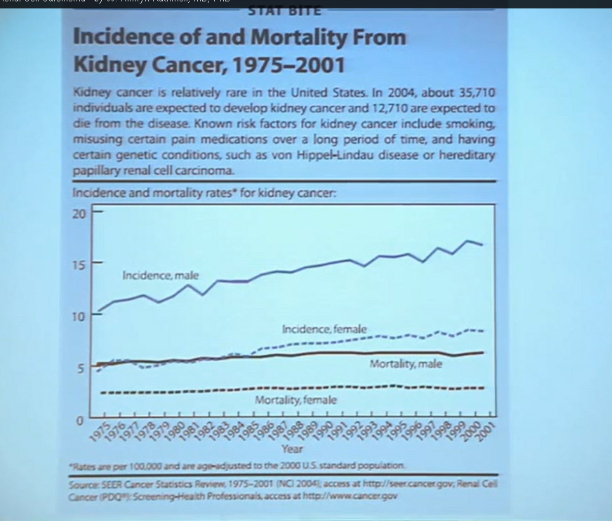
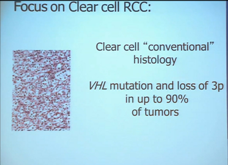
First, kidney cancer, like pancreatic cancer, has been on the rise. This is a somewhat dated slide, dating back to the 70s. We have seen a steady increase in this cancer. Although it was originally characterized as a rare tumor type, it is not really anymore.This talk will focus on one subtype of kidney cancer, that is, clear cell histology renal cell carcinoma. This is a histology slide showing why it is called clear cytoplasm
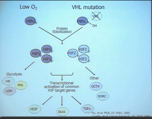
This tumor is characterized by particular mutation. That is the Von Hippel Landau gene, coordinate the loss of 3p, (a chromosome) where VHL is housed. We see these in mutations and loss of 3p which house other tumor suppressors as well, in up to 90% of these tumors. Based on this strong correlation between clear cell renal cell carcinoma and the VHL mutation, a tumor type. is a very distinct paradigm in which VHL loss causes upregulation of hypoxia inducible factors (HIF). These tumors are characterized by loss of high loss of these HIF factors. These are transcription factors that normally allow cells to respond to low levels of oxygen by turning on a repertoire of genes that allow them to bring in new blood vessels, to shift their metabolic properties, to migrate away, to promote survival, and to de-differentiate. That is a perfect storm for kidney cancer, in some respects.
Targeted Agents in Use
Because this cancer has highly nonresponsive to typical chemotherapy, there’s much effort in recent years to develop targeted agents. These targeted agents to date all focus on this well-known pathway in clear cell type renal cell carcinoma. Most of the agents focus far down on this pathway, including that of receptors of VEGF and PDGF. They are tyrosine kinase inhibitors, effective at reducing the tumor angiogenic profile and can be quite effective at reducing the bulk of these diseases. Other drugs similarly target this pathway, for example, targeting features of the tumor that enable HIF to be stabilized such as that in the mTOR pathways. Temsirolimus and Everolimus are approved for use. There are in-developments drugs for targeting MET, which is another mutation that can occur in this cancer, similarly increases HIF levels.
But the reality of treating kidney cancer is that the available drugs that we have do not produce complete responses. We only work in the arena of minimal response and partial response. The extent of response that a patient gets is unpredictable. The duration is also unpredictable and the toxicity is also unpredictable. For drugs we expect them to be effective on average 1 to 2 years, this is chronic therapy, very expensive, and it’s dominated by effects that are substantially detrimental to quality-of-life—fatigue, rash, diarrhea, as well as laboratory abnormalities that indicate damage to the liver or elevations of glucose and cholesterol.
PART I: Clear Cell Renal Cell Carcinoma, Molecular and Genetic Contributions to
INTER–Tumoral Heterogeneity.
With that, I will talk about various molecular probes that we use to understand some of the diversity or the heterogeneity of these tumors across the clear cell renal cell carcinoma spectrum. Before I really dive into clear cell renal cell carcinoma, I need to point out that there are other histologies with this tumor as well. So when we say kidney cancer, we’re talking about a big spectrum. Clear cell renal cell carcinoma, we are talking about those tied to Von Hippel Lindau disease and loss of 3p and it is about 70% of all cases we encounter in cancers of the kidney. But there are also other types. Papillary type renal carcinoma, chromophobe, benign tumor—oncocytoma, a translocation form and some very rare. With these types of tumors we have very little in terms of knowledge of how to treat these patients. Their genetics are highly distinct from clear cell renal cell carcinoma. So someday in the future, we will understand not only how to treat not only our clear cell carcinoma patients, but how to use effective molecular information to target these cancers as well.
Clear cell carcinoma is well know to be molecularly heterogeneous for some time. This is a gene expression profile. We’ve already seen heat maps from several of these other talks, looking at gene expression profiles. And as you can see the gene expression profiles across a large selection of tumors here, suggests there are great areas of variability–at least two and as many as five groups, based upon gene expression purely.
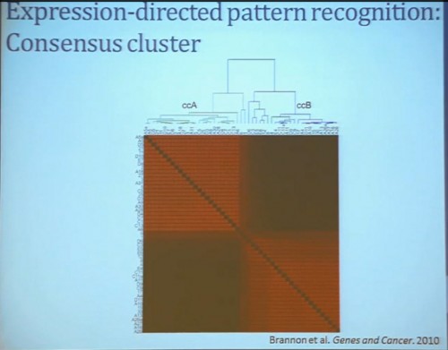
Pattern Recognition Profile to Find Subtypes
Our group undertook at the time developing a pattern recognition profile which is now fairly routine use. To try to see with a more robust computational strategy what subtypes we could really identify, that we could really pen down and understand with genetic profiles. 8aWe found two. For lack of better knowledge, we are calling clear cell A and clear cell B, ccA and ccB. These are very distinct biologically, and when we look at these tumors in terms of their outcome, they also have significant prognostic relevance–with the ccA tumors in this original cohort having a median survival of 103 months, compared to the 24 months for ccB tumors.
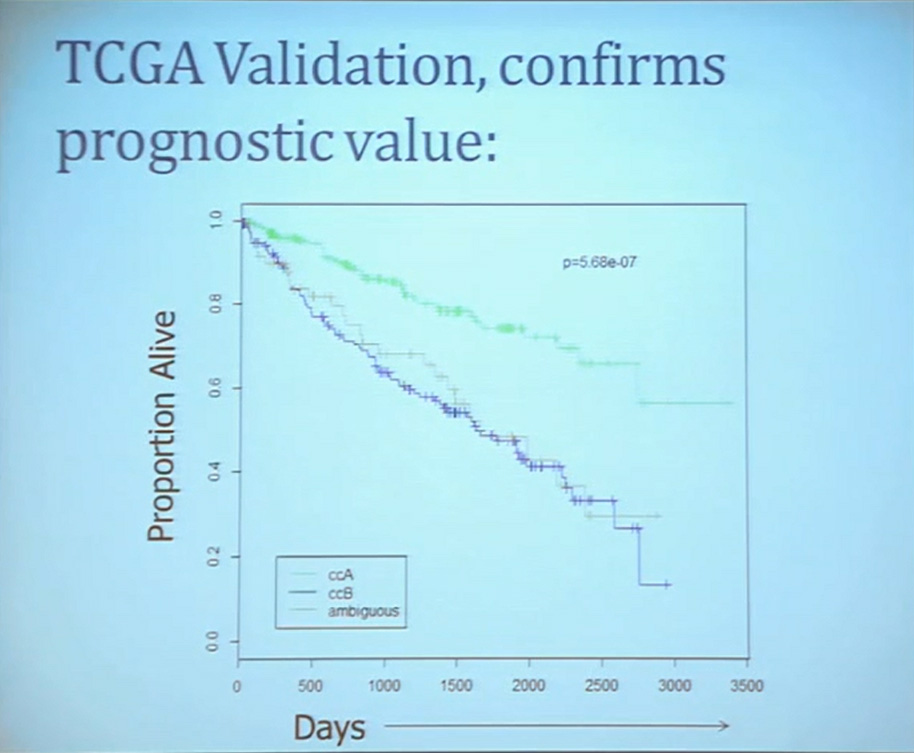
The TCGA which is been discussed here in many of the previous talks is a great source of validation. We assigned these tumors to clear cell A and cc B groups subtypes, validating our previous results with the clear cell A tumors having much better survival profile than those ccB tumors.
This classification scheme, which is based 120-130 gene signatures classified robust subdivisions of clear cell type renal cell can be applied with a small number of genes on individual tumors and is independently associated disease-specific overall survival, making it a valuable prognostic biomarker.
PART II: Rare Variant Groups
We use these profile tools to understand the rare variants. This is still in the clear cell renal cell carcinoma arena, but when we took a very large group of compiled tumors; this was a meta-analysis of 500 tumors, all histologically defined as clear cell type renal cell carcinoma, and we applied our expression pattern recognition algorithm. We asked for two groups and we found two and they correlated with our ccA and ccB, but when we ask for three groups, we can find a small group that now filters out. Now that we have power in numbers to identify what we called Cluster 3. What is in Cluster 3?
Cluster 3, as we’ve said, is histologically defined as clear cell renal cell carcinomas. But we look to the genetic expression profiles, it’s very different, particularly with regard to metabolic properties. We see upregulation of genes that are involved in mitochondrial regulation and oxidative phosphorylation, suggesting a striking difference in the way these tumors likely regulate metabolism.
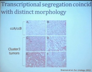
In addition, and now these are tumors that we can not go back and genotype for VHL mutation, for loss of chromosome 3p, but the loss VHL regulation leads to characteristic changes in the gene expression profile. So when we use the gene expression changes to predict whether these tumors have an intact VHL or a mutant type VHL. The wild type VHLS signature shown here is shown in purple. You can see that these purple tumors, the wild type VHL tumors all tightly cluster with Cluster 3. These are probably not clear cell renal cell carcinomas although many, pathologists call them that. So we pulled them all out so, asking, “DO they look a little bit different?” My graduate student, who did this work, came right away and said “There’s something funky about these clear cell tumors that we call Cluster 3.”
As you can see, these are clear cell A and a clear cell B tumors, but they all have the clear cytoplasm and really, what we are seeing, is that they are not distinct histologically, although they are very different molecularly. And as I have shown, they have a very different prognostic outcome. The Cluster 3 tumors; although the cells themselves might have clear cell cytoplasm that gave them the clear cell histology designation, they have a very different pattern of organization—with a papillary type of feature. So what we think it would be identified as is a new rare variant of clear cell renal cell carcinoma.
Simultaneously another group of pathologists identified, that the pathologists call clear cell papillary carcinoma. That suggests that we need to take a very great care as we treat these patients. What we have is clear cell type renal cell carcinoma, most of which are VHL-mutated, and we do have clear cell A and clear cell B. These are the tumors we should be treating with the drugs with identified, based on the effect of the pathway that is activated by loss of VHL. But clear cell papillary renal cell carcinomas probably won’t react very well, as they are VHL-wild type. Just like papillary renal cell carcinomas don’t react well either.
To summarize this section, clear cell renal carcinoma can be separated into ccA and ccB groups, based on transcript profiling, but further clustering can identify highly biologically dissimilar subtypes within the clear cell group, and that subtyping can convey a biological distinction which is a valuable tool for prognostic evaluation, and a likely cause of poor responses to some therapy.
Part III; Using Clinical Trials to Understand Biological Relationships to Response to Therapy
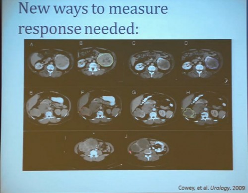
As my title indicated, we also refer to clinical trials to help us understand renal cell carcinoma a bit more. A clinical trial we completed some years ago, LCCC0603, was in neoadjuvant trial that looked at the treatment of renal tumors with sorafenib. Patients were identified as having renal tumor and underwent CT scans for basic size, description and PET scan, and then were treated with sorafenib. It is the first generation VEGF receptor tyrosine kinase inhibitor for 4 to 8 weeks, and then underwent post treatment CT scan, PET scan and a nephrectomy. We are going to look at radiographic indicators of response, rather than molecular indicators.
Waterfall Graph with Response at 30% as goal
First standard RECIST criteria do show that we do see partial responses. Again, there were no complete responses. Many had subpartial or minimal or some partial responses. Their tumors shrank, but most did not meet the standard criteria of 30% decrease in one longest size. Some tumors actually grew.
Now what we realized as we looked at these tumors, is that we probably need new ways to describe response. The standard RECIST criterion response is just based on longest diameter and measuring this in comparison, after treatment..
I will use this patient is an example. (References I and J, in the lowest row). Here is a pretreatment; we have a very large renal tumor. Post treatment, the tumor was still large, but it measures slightly larger than it had been before. But if you look at this tumor, it is very different. The central area of this tumor is now very dark, indicating necrosis—is what we think. But we took these tumors out, we could confirm that these dark areas were indeed necrotic. So we developed a new way to try to quantify the area of the tumor that is actually killed in response with this treatment.
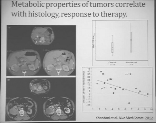
Similarly we were doing PET scans on these patients, and we were doing this because we’re trying to understand how the metabolic properties of these tumors might indicate how these tumors were likely to respond to this treatment. We see, and have known, that are some tumors which are very dim on FTG PET. This is a tumor; (Smallest of upper images) you can see that here is very visible on the PET scan. It doesn’t take up any FTG. So this tumor has the metabolic profile that is not dependent upon uptake of glucose. Others; this tumor (Smallest of lower picture), for example, have regional areas that are can be very high in terms of FTG uptake. When we looked at these tumors, we discovered first that non-clear histology tumors were much more likely to have high levels of FTG uptake. So, metabolically active tumors more likely in the (correction) non-clear cell group, probably the papillary, the chromophobes and the papillary clear cell types, than the clear cell group. Secondly. we discovered that the correlation between FTG uptake and response looked somewhat different than we might have expected. We might have expected the most metabolically active tumors would be those that would response better to anti-angiogenic agent. But the opposite was true. The best are those that had very low levels of uptake FTG uptake. We are still trying to understand exactly what that means. Certainly that means that those clear cell tumors are the ones more like to respond, activity, what we have known, but those are the ones with the lower level FTG activity. But we continue to try the metabolic properties of the tumors that make theme different more likely to respond.
That leads to our next clinical trial. This is now ongoing. This is LCCC1028. It is a neoadjuvent clinical trial using the newest generation—well, they are coming out so fast that it’s the not the newest, but the next to newest VEGF receptor tyrosine kinase inhibitor. They are now getting PET scans and a biopsy to confirm in fact that they would be clear cell renal cell carcinoma, and also to allow us to do molecular studies that directly measure their metabolic activity and other effects. They’re being treated for eight weeks with another CT scan, undergoing nephrectomy. We will then be able to look at clear cell variant histology. They will all be clear cell going in, but there may be variants included–as well as looking at their VHL mutation and their other mutational status, their transcript profile, in particular the clear cell A and clear cell B group and other protein expression signatures.
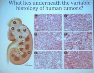
This, of course, is known for all tumors, but if you sample in multiple different places, the histology will look different and in effect, the grade can look different depending on where you are sample.
What does that mean molecularly? Well, a group at the Sanger Center published on a small number of tumors. When they sequenced these tumors, they found while there are some mutations that are ubiquitous, meaning the mutation is found in all samples across the primary tumor and the metastatic tumors, that there are mutations that are private. There are mutations that are common only among the primary tumors and there are mutations that are common only in the metastases, and there are a lot mutations that are unique to the individual sample. This makes a whole new level of complication as we moved toward personalized therapy, in particular therapy that is based on biopsy metrics.
This group also looked at our clear cell A and B subtypes. And what they saw, when they looked at six samples from the primary tumors, was that in five of those samples, the gene signature indicated that these would be clear cell B type tumors. So depending on your glass half-full/glass half empty: The glass half full version of this, that five out of six times, they would pick that the patient would have poor outcomes. This patient has metastatic disease, so it fact, that is true. The glass half empty would be that one out of six times, he would pick wrong. This patient would have been indicated to have clear cell type A tumor, and you might have predicted that this patient would do well, when in fact that would be wrong. So what helps us understand the limitation of this test. It also gives us the opportunity to understand a little bit more about these tumors.
(Footnote reads: BRIC Funded Grants LCCC1213) So for the future, a trial (LCCC1213) that we have really just initiated is uniting some of these imaging observations we have made with genetics.
We are taking in patients. This is patient number one. Patient number two just has his MRI last week. and doing an MR in coordination with the PET scan so we can get the detailed look at these patients’ tissue perfusions, vascularity and the density of these tumors, as well as regional areas at the FTG uptake and sample according to the map that is created by the imaging, as well as samples that are collected, based on what we see grossly in the tumor. Here you can see a sample that we collected from a tumor region that is highly distinct from the mostly more pale yellow regions of tumor.
This is just begun, so I cannot tell how well were going to get to correlate the gene expression and genetic underpinnings, and what we see in the tumor and what we see in the MR / PET. But it will help us to move forward.
To summarize are multiple ways for RCC to diverge. The subsets can enrich tumor sets for clinical and genetic features, and a multiplatform approach that with genetics, molecular biology and imaging techniques will give us man ways to tackle a surprisingly very heterogeneous disease.
Once a patient can stop asking why cancer happened to him/her, the next question is the fundamental version of the many questions that the researchers ask– “What is cancer really? and “Why can’t they just stop it?”.
The scary part of cancer is that it seems so insidious. Cut it out. Burn it out. Zap or freeze it out. Why doesn’t that work, at least for the solid tumors? Getting rid of the blood cancers, like leukemia and lymphomas seem more difficult, less obvious, but it was really the cases of leukemia which first responded to treatments.
Without going into volumes of discussion about cell and molecular biology (you are safe from that with me), just understand that cells go wild, left to their own with the family checkbook, an endless liquor cabinet, permanent pizza delivery, car keys, disguises, blind neighbors, a fancy cloning machine, and the police on strike. You get the picture. Now a more formal explanation.
Cells are supposed to do their respective duties and then die. That process is called apoptosis. You know that your scabs don’t keeping growing, but cancer cells lose the “time to die” signal.
Foreign bodies are supposed to be cleared by the immune system, and what is more foreign that cancer? However, cancer cells manage to evade immune destruction. And while doing that, they can also evade the growth suppressors, the immune brakes which would otherwise slow and prevent excess growth.
While cutting the brake lines to growth, they can also change the regulating signals for growth (think scars and healing), so those signals all left in permanent “ON” position. No brakes and an open throttle with a very full tank of fuel. To top that, they reprogram the nutrition or energy metabolism to keep the fuel of growth alive.
Liking the growth, the cancer cells override signals that naturally limit the times a cell can divide, creating endless replication instead. With the endless replication, the chances for mistakes, or genetic mutations increase, which can mean changes from the original cancer cells. Sheesh, not only alien cells, but aliens cells making alien-er cells!
To keep tumors growing, cancer cells send out signals to create blood vessels or angiogenesis when tumors outgrowth the local nutrient sources. Running out of room for all these many cells, billions and billions, the cells break down the lining of blood vessels and the lymphatic system to search out new locations, spreading and metastasizing away from the original cancer. Quite naturally, they also provide support to those tumors through inflammation-related factors, mimicking the way that the immune system responds to any injury.
Since a healthy being grows, fights off infections, responds to an allergy or heals after an injury, usually with little support, those healthy responses are amazingly efficient and interactive. Complex cellular, molecular and chemical actions are occurring all the time, and with aging, some genetic dispositions, the harmful things we ingest or do to ourselves, it is no wonder that a few things go on without our noticing it. But when those few things are not noticed by the immune system, slipping into a growth phase, a cancer can begin.
Estimates of the numbers of cells in the human body are calculated from 10 trillion to 100 trillion, so if the occasional cell goes rogue, what’s the problem? When all things are working well, there will be no problem. But when the tiniest cancer is visible with a CT scan, perhaps 1/8 inch, it will have millions of cells. Not all tiny cancer tumors are dangerous. Not all become aggressive. Digging around to cut out a tiny tumor creates plenty of opportunity for infection, for expense and emotional anguish. But does that mean that a “Cancer!” has been prevented. Or would the person and his natural immune system have lived in complete tranquility with his cancer until the end of his days?

 Two arms/groups: BLUE arm with surgery and monitoring by the trial team, the standard of care; the RED arm with medication before to surgery, followed by more after the surgery.


 QED
QED