Surgical Management of Locally Advanced Renal Cell Carcinoma
Next is the management of more advanced disease, in patients who present with tumor thrombi, nodal metastases, adjacent organ invasion and renal fossa recurrence.
The first is venous tumor thrombi. This is one of the biggest operations that I do, as about 15% present with venous thrombi from their kidney cancer. These can be huge. This can be very complicated, as these thrombi can go up into the heart. We then have to put these patients on by-pass, open up their chests, stop their hearts and put them on a bypass, so we can take the thrombus out. We use TransEsophagealEchocardiogophy to monitor of the thrombus, to make sure that the thrombus does not break off.
This is a series that we recently published at MD Anderson, 605 patients with venous involvement, median age of 60 years, follow up of 24 months. 45% had no evidence of metastatic disease; conversely 55% obviously had advanced disease. The more advanced stage, the higher the risk of metastatic disease.
These are big operations; median blood loss is a liter and length of time is 3 hours, with complications inthe first 30 days for 25% of patients. Patients stay in hospital about six days. But even with venous involvement up to the heart, in the absence of metastatic disease, there is a median survival of five years, so even these patients can be cured with surgery alone. But once they demonstrate evidence metastatic disease, survival drops off dramatically.
Predictors of Overall Survival Slide
We looked at a variety of different parameters, won’t bore you with all the details. We looked to see what predictor survival for these patients and this is what we found.
Those patients who had clear cell histology actually had a more favorable outcome than those with non-clear cell. Those who had advanced grade, sarcomatoid de-differentiation, those who had peri-nephrectic fat, nodal metastases, distant metastases, all were associated with a more adverse outcome than patients with venous thrombus involvement.
Interesting was in our studies, not shown elsewhere in the literature, is that the height of the thrombus in the vena cava did not seem to matter. In other words if it was only in the renal vein, versus in the vena cava and that is at odds with the literature that is out there. It only seemed to matter when it went up into the heart, the red line at the bottom and with that, we saw a decrease in overall survival.
Tumor Thrombus and Survival
Our series are in concert with others, in regards to survival in the absence of metastatic disease that patients can have a long term durable survival, with about 60% with five year disease-free survival
Microscopic Positive Vein Margins Associated with Increased Local Recurrence & Metastatic Progression
One of the other things we noticed from our series that made me wake up was the concept of the vein margin. Of 270 patients, who had no evidence of metastatic disease, almost 20% of patients had cancer present at the margin of resection. When we resect the vena cava, we are obviously limited in how much we can take, unless we are doing a reconstruction, which is really fraught with complication.
(associated with increased metastatic progression.
But those with a positive margins, meaning they had cancer sitting at the edge of resection, where we cut the vein, had a greater risk of local recurrence, a higher Fuhrman grade. Those patients had a worse outcome, not surprisingly.
So now when we do these surgeries, we send the resection for a frozen section to be sure there is no cancer at the margin. If there is cancer at the margins, then we will do a reconstruction to try to reconstruct the vena cava to try to eliminate all the cancer.
Also in the literature that is out there that is a bit at odd with our series; here they noted that patients who had only renal vein involvement had significantly better outcome than those who had IVC (Inferior Vena Cava) involvement. There is some data that suggests that the height of the tumor in the vein is somehow related to outcome but that is not what we are seeing in our patients.
In this series, IVC (inferior vena cava) wall invasion, tumor size, fat invasion, nodal metastases, and distant metastases all were associated with an adverse outcome in patients with venous tumor thrombi.”
Dr. Wood continues his discussion of surgical management of “Locally Advanced Renal Cell Carcinoma” with Budd-Chiari Syndrome in Part 3.
Dr. Christopher Wood, of MD Anderson Cancer Center lectured at an April 2012t KCA patient conference on this critical subject. It is a technical discussion, highly important for the patient whose kidney cancer is not longer consider “small and curable” by surgery alone. For greater ease in following this, I have posted this in four segments. My comments will follow in a separate blogs. My hope is that the information is meaningful, so that a patient can learn enough to have a discussion with his/her doctor. Print out the information you have read and give it to the doctor to start that discussion. Some slides were principally text, so I have recreated some to keep the file size down; anything which was not readily converted to text remains as the best available slide.
Consider saying to your doctor, “Here’s what I have been learning, doctor. How does that fit into our plan for my treatment?” That tells your doctor know you are willing to learn and to listen, and to take an active role in your treatment decisions. Unless your doctor deals daily with many kidney cancer patients, he is unlikely to be able to keep up with all the information, ever changing, and ever more complex, about your disease. If the doctor is not willing to listen and learn from the leaders in the field…find another doctor.
Peggy: RCC Patient, Stage IV mRCC 2004; healthy today, thanks to HD IL2
“Outcomes for Patients with Locally Advanced Renal Cell Carcinoma”
Dr. Wood begins: “My first lecture is “The Management of Locally Advanced Renal Cell Carcinoma”. Slide 1
Stage is the most important predictor of outcome: the more advanced the stage, the greater risk that the tumor has spread, with distant metastases, making the disease incurable.
This is the staging system we use in 2012, where we stage tumors, assess regional lymph node involvement and look for evidence of metastatic disease. People get hung up on their staging, but this allows doctors to communicate about patients in the same language about how advanced one tumor is. With increasing stage, there is a more locally advanced tumor or metastatic disease.
Let us start with definitions. (Reads slides 2 & 3)
Adjuvant Therapy means some form of therapy– chemo, radiation, vaccine, whatever–after complete surgical tumor resection with the idea to decrease the risk of recurrence of disease. The benefit is that the patient has already had surgery before getting additional therapy, but the downside is that many of those patients may have been cured by the surgery and they may get treatment they don’t really need.
Neoadjuvant therapy is taking some form of therapy, whether chemotherapy, radiation, vaccine, etc. prior to surgery to the primary tumor in hopes the tumor may decrease in size, and decrease the risk of recurrence. The benefit is it may allow the tumor to shrink and make surgery easier. The downside is that the therapy may not be effective and not inhibit metastases, and the primary tumor would not regress, but progress during therapy.
Slide 4
Effective adjuvant or neoadjuvant therapy does not exist for kidney cancer in 2012
Any therapy must be developed in the context of a clinical trial setting, and including a placebo. The only way we make advances is to test what we do now with any advance in the future. And what we now know, as I said, is nothing.Patients have a hard time, with placebo trials. If you participate in such a trial, the treatment you are getting may not be good and the placebo arm, not so bad.
Slide 5 Who should get these therapies? Why not give them to everyone? Anyone taking the targeted therapies today knows the toxicity is significant, and you may treat a significant number of people who really don’t need the therapy. Then if nothing works, why not give it to anyone? Well, we are never going to make advances that way, so it is important that we continue to do research and focus on those who are highest risk for relapse. Should we give it only to those at a highest risk for recurrence? The difficulty how do we define risk, how high is high? So it’s not really clear.
Adjuvant Therapy: 2012
A variety of trials have been performed. Many patients have participated. They include radiation, embolization, energy ablation, a variety of different hormonal therapies, immunotherapies with interferon and interleukin 2, all having been used in an adjuvant setting. There have been a variety of vaccines preparations and we did a Phase III trial of thalidomide trial here. To date, not one ofthese therapies has shown benefit in the adjuvant setting. In fact, many of the patients on the treatment arms did worse than those who were not treated.
Slide 7 What about targeted therapies? That is also the great unknown where things stand with targeted therapies in the adjuvant setting.
Since 2006 there have been seven new agents against kidney cancer. It’s been a revolution. And to be honest, many have benefited from that advance. How do we use these agents in the context of adjuvant therapy?
Slide 8 There is a variety of trials recently completed or in accrual, ongoing. Tthe ARISER Trial used an antibody called G250 against Carbonic Anhydrase IX, and patients were randomized to get either antibody or placebo. This trial completed accrual many years ago, in fact, and we are still awaiting results which leads me to believe that is probably going to be a negative trial.
But this same agent has recently shown promise for use in PET scans for kidney cancer. You can use this as an imaging agent. The patient is infused with the antibody and linked to item 125, and it will show up on x ray, and may potentially detect micro metastases not visible on CT scans. This is actually undergoing FDA approval.
Slide 10
This is the ASSURE trial we conducted at MD Anderson. It is a randomized, double-blind phase III trial of Sunitinib vs Sorafenib vs Placebo. Patients underwent surgery, then were treated with these agents for 1 year. This trial was completed accrual last September (2011) and we are now waiting for the trial to mature to see whether these agents have any benefit in the adjuvant setting.
Slide 11
One thing that we did learn from this trial is that tolerance for the toxicity associated with targeted therapy in the adjuvant setting is not the same as in the metastatic setting. In this trial 41% of patients stopped therapy early, not because disease returned, or because they finished, but because of toxicity. I think it comes down to an individual assessment of the risks. If I told you that you have a 20% risk of your cancer coming back, versus your 70% chance of your cancer coming back and you are miserable on this therapy, all of a sudden 20% doesn’t look so bad.
My concern about this, because at the end of the day, if this trial matures and it is negative, will it be negative because the agents did not work, or because the patients could not tolerate the side effects. And too many patients stopped the trial early or had dose reductions. I’m afraid it is not going to be interpretable.
Slide 12 This the S-TRAC trial, sponsored by Pfizer, recently completed accrual. It’s randomized between Sunitinib with a placebo for one year. It is estimated results will come out in 2017. That’s the other problem with these trials. Are the agents we are testing now, will they even be relevant in 2017? No one knows.
Slide 13/12
There has been a real problem in accruing to this trial. In fact, we can’t even keep patients on Sorafenib for three years in treatment, never mind adjuvant setting.
Slide 13
This is a trial going on in the US, sponsored by Glaxo-Smith-Kline. This is called the PROTECT trial. Patients are randomized to Pazopanib (Votrient) or placebo for one year. This is primarily open to clear cell patients, here at MD Anderson.
SLIDE 15 A
This is the EVEREST trial, sponsored by the SWOG, going on around the US, with patients randomized to one year of Everolimus (Afinitor) or one year of placebo.
You can see that clinical research is actively ongoing to identify if these agents work in the adjuvant setting. But it is going to take more time to understand from these trials before we know if this is applicable in these setting or as with the other agents, if they remain ineffective.
Slide 16
One other concept we are testing at MD Anderson that I will talk about a bit more in the next talk is a neoadjuvant therapy, testing Axitinib in the neoadjuvant settings. Some in the audience have been on this trial, where patients receive Axitinib for three months and then undergo nephrectomy.
I would like to applaud the patients who participated in this trial. Like to give you an idea how this went the first time we enrolled a patient. Patient comes in with a tumor, curative with surgery, and we say, “We’d like to give you this agent, we don’t know if it will work. In fact, your tumor may grow and metastasize while you are getting this agent. If we took you to surgery tomorrow, we could probably cure you. What do you say?” It was really amazing. The first patient who enrolled in this trial was very brave. But since we have been able to show activity with this agent, enrollment has picked up significantly.”
Dr. Christopher Wood ends the first part of this lecture, with the next section about “The Surgical Management of Locally Advanced Renal Cell Carcinoma.”
The Small Incidental Renal Mass: Treatment Options
I have summarized and edited presentations from an April 2012 Kidney Cancer Association Patient Conference so patients can read and study them.
The original presentations can be found on YouTube offered by the KCA.
Poor resolution slides have been reconstructed
Treatment Options for the Small Incidental Renal Mass
After a brief welcome and expression of appreciation for the KCA, Dr. Karam begins:
“Currently, a small renal mass is defined as smaller than 3 or 4 cm, localized to the kidney, not invasive to other organs. This is a consensus and these definitions can change. Of all Stage I kidney tumors, 43 % are less than 3cm in size. (Some Stage I kidney cancers can be larger than 3cm; other treatments may be more suitable.)
I will discuss three options with patients who present with these types of lesions. The first is active surveillance, the least invasive, the next is energy ablation, and the third is surgical removal. Each will be discussed in the sequence.
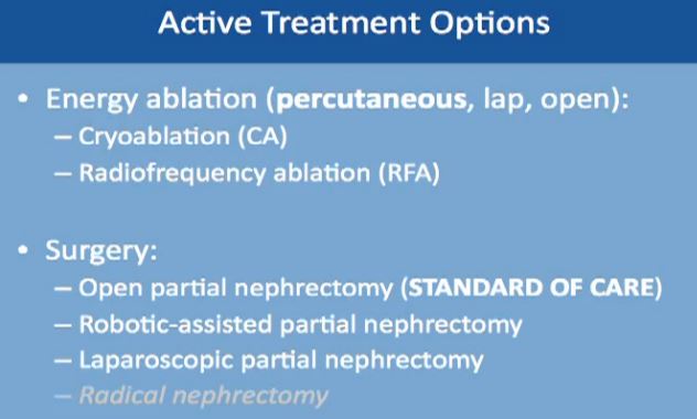
First “Active Treatment Options” is energy ablation. This can be done through the skin, percutaneously, laporascopically, or be done “open”. Energy ablation includes two categories, Cryoablation (CA)–freezing the tumor, done laporascopically or through the skin. Alternatively is Radiofrequency Ablation (RFA), typically done percutaneously, or through the skin.
The second major category of active treatment is surgery, the standard of care being an open partial nephrectomy. That removes just the tumor, leaving most of the kidney. It is now more often being done as a robotically assisted partial nephrectomy. Another option is to do it laporascopically, with minimal incisions. Radical nephrectomy, complete removal of the kidney along with the tumor is the lowest on the list. It is the least favorable option, as we try to save the kidney, wherever possible.
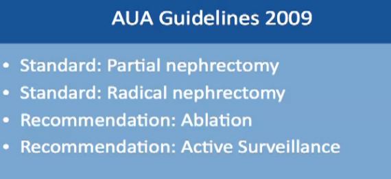
AUA (American Urological Association) Guidelines have published some guidelines, based on multiple studies and experts. The standard of care for treatment of small kidney masses is partial nephrectomy. A radical nephrectomy is used only if the mass cannot be removed by partial nephrectomy. A recommendation could be ablation or active surveillance. This will be tailored to individual needs of the patient and the kidney, as to the location of the mass and its characteristics. No one recommendation fits all patients, and each must be discussed and individually tailored.
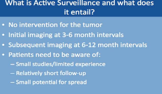
Next is active surveillance. We don’t use “watchful waiting”, but “Active Surveillance”. We do not to tell someone to go home with your small mass as we don’t need to do anything about it. On the contrary, we have to actively bring the patient to the clinic for CT scans.
What is active surveillance and what does it entail? That means doing no interventions, no surgery, and no procedures that are invasive–active surveillance. Initially we would do imaging at 3-6 months intervals in the first year, subsequently imaging at 6-12 months. There are no rigid guidelines at this point. I always tell my patients to be aware that we have only had small studies with active surveillance, and with short follow-up of 2-3 years. There is a very small potential for spread or metastases while we are doing active surveillance, so every patient has to be informed about all of this.
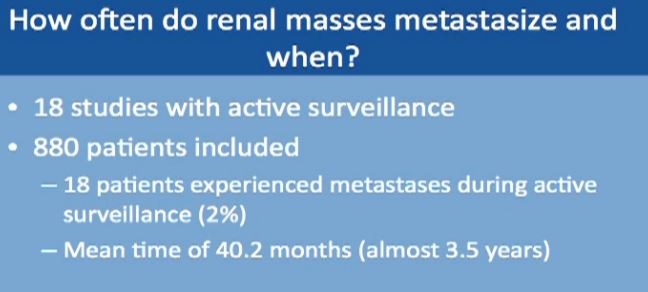
How often do small masses spread or metastasize? When does it seem to happen? This is a meta analysis of data from 18 studies with almost 900 patients. In only 18 patients of 880 did metastases occur while on active surveillance, a rate of about 2%. This spread occurred almost 3 ½ years after initiation of surveillance, so it does not occur in the first 3-6 months, more on average about 3 ½ years.
“How fast does a mass actually grow?” We know that 23% do not grow at all within 2 years or so. The rest do grow at the growth rate shown.Comparing the growth rate of those who had spread or metastases with those who had none, only 18 patients had spread. The group without spread was typically younger (66 versus 75), with a smaller mass at the initial presentation, 2cm versus 3 cm. The final size in centimeters of those who had spread, those 18 patients, was about 5.9cm. The tumor was quite large at the time when the metastases occurred, and here we started at a size of 3.1 cm.
The growth rate was about 2mm per year for the patients who did not have spread, and about 6mm per year for patients who did have spread. The time on active surveillance was about 2 ½ years.
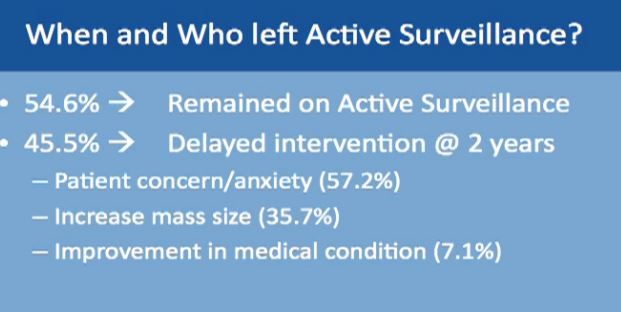
Of these patients on active surveillance, what happened to them, did they all continue after active surveillance? Over half did remain on active surveillance, over a period of 2-3 years. Fewer than half had delayed intervention. They might have gone to ablation or surgery, and this is over a period of 2-3 years. Others had “delayed intervention, with patients having ablation or surgery on average about two years after starting active surveillance.
The common reason for moving from surveillance to intervention was typically patient concern about the growing mass—patient anxiety. The next reason was enlargement of the mass and the third one was that a patient had some bad medical problem initially not related to the mass. When that problem was solved, and became a good surgical candidate and they are switched to surgery.
Energy Ablation: Radiofrequency
Next is Energy Ablation, a more invasive category which includes radiofrequency and cryoablation.
How does radio frequency ablation work? I usually tell my patients that is basically frying the tumors. The waves from radiofrequency cause friction and movement in the water molecules. This produces heat and destroys the tumor. Cell death occurs in about five minutes after exposure to the RF, with the temperature over 50 degrees Celsius )122 Fahrenheit. This can be monitored during the procedure. We have to achieve temperatures up to 100 (212 F)degrees in order to effectively destroy the tumor with this procedure.
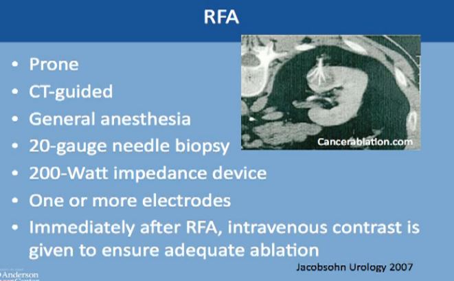
This is a CT scan of the vertebral column and the patient is laying flat on this tummy. This is the kidney, this is the mass, and is CT guided. The interventional radiologist usually puts this thick needle with these tines, typically done under general anesthesia. Though we can do it very infrequently under conscious sedation, but general anesthesia is preferred. A biopsy is typically done at that time and one or more electrodes are inserted. Immediately after the procedure iss done, we do a CT to see that there isn’t any bleeding and to make sure this was treated adequately.
Energy Ablation: Cryolablation
(This is a video which can be seen on the Youtube video.)
The next category of energy ablation is cryoablation or freezing the tumor. This is a picture during laparascopic surgery. You can see them freezing the tumor. This is the kidney here, the tumor here. The tumor here has been marked; the electrodes that go into the tumor, freeze the tumor and form an ice ball.
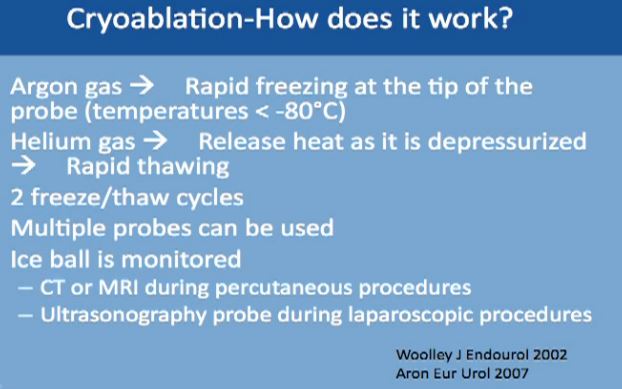
We use two gases for this procedure. The first is argon, which causes rapid freezing at the tip of the probes, with the temperature dropping to less than 80 degrees Celsius. The second gas is helium, which releases heat as it depressurizes, and it causes very fast thawing of the tumor. This typically repeated twice, two cycles of freezing and thawing. Multiple probes can be used to achieve tumor ablation. With this technique the ice ball can be monitored and you can see the progress with time. This can be monitored by CT or MRI, done percutaneously under guidance, or with an ultrasound when you are doing it laporascopically. or with an ultrasound when you are doing it laporascopically.
Cryoablation—How Does It work?
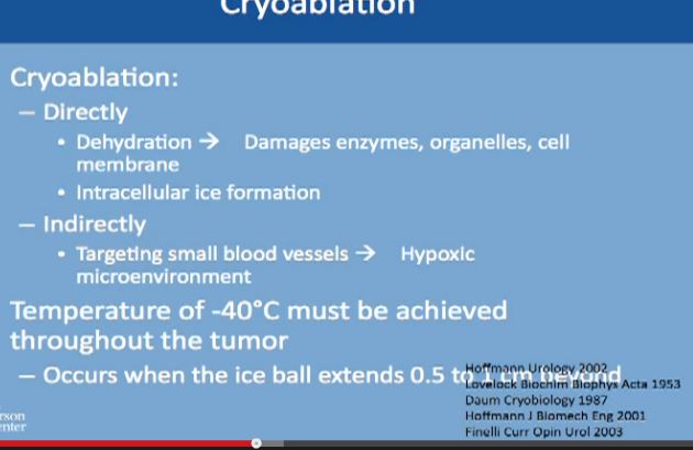
The Cyroablation acts directly on the tumor cells. It dehydrates the cancer cells, removes all the water from them, which causes damage to the enzymes which are needed for survival by the cells, the organelles and the cell membranes. It also causes the formation of ice inside the cell and kills the cell. But it can also act indirectly, as it targets the cells directly, cutting off nutrition needed for growth. In order to achieve this, very low temperature have to be reached, and to achieve it homogeneously for all of the tumor, the ice ball must extend ½ to 1 cm beyond the margin of tumor to make sure it is completely frozen.
Cryoablation
As to follow up, how do we know it is still working? There will be routine visits to the clinic with medical history, physical exams, chest imaging which is either with conventional radiography, CT and routine blood work.
Follow Up
To follow up on the tumor itself, we typically do a CT or MRI with IV contrast to visualize the tumor. The first follow up is at 6 weeks after the ablation to make sure the ablation was complete and successful and then at 6, 12, 18 and 24 months after ablation.
It does not mean that follow up will end at 24 months. The patient continues with scans for life, routinely, at least once a year after deletion or the treated tumor is stable. So imaging has to continue for years, as we don’t have long term studies with these technologies.
We tend to do more biopsies if the mass is not shrinking well enough, or if it is taking contrast, or it doesn’t look right one year. If the mass is not looking right, we go ahead and do the biopsy to make sure there isn’t any recurrence.
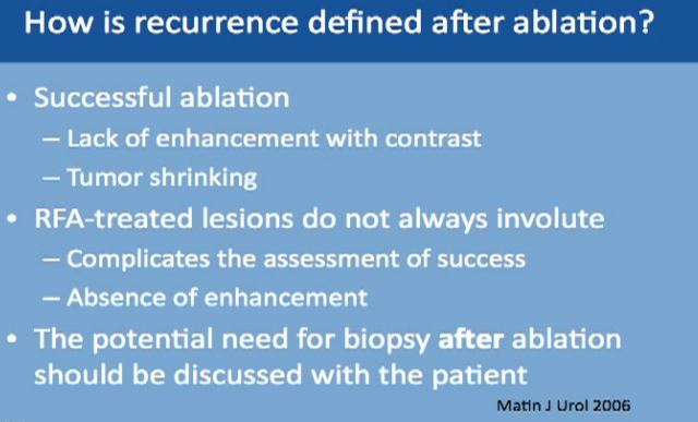
Recurrence defined after ablation can be tricky because the mass is still there. A successful ablation is defined as lack of enhancement of the tumor with the use of contrast. That is why is it very important for the patient to receive and tolerate contrast IV contrast for follow up after ablation. With cryoablation, typically you see tumors shrinking. With Radio Frequency, it doesn’t always happen. That is what makes it trickier, so we have to do biopsies for patients after treatment. And again, the tumor does not always shrink; more typically it does with the cryoablations. Again, radio frequency ablation lesions don’t always shrink, so absence of enhancement is what we look for which is not always ideal.
Now we always discuss with our patients the potential need for biopsy after ablation so this may be expected, though is not routine. Thus, the patient has to be aware of this. We don’t do it routinely, but it is a possibility.
What can predict who is going to have a successful ablation? Tumor size less than 3 or 4 cms is a very good indicator that we will be able to successfully ablate the tumor. If the tumor is exophytic, that the tumor is not deep into the kidney, this is a good indicator. If the tumor is peripheral or not central, that means that the tumor is away from the artery and the vein of the kidney. This is also a very good indicator that we can successfully treat it with ablation.
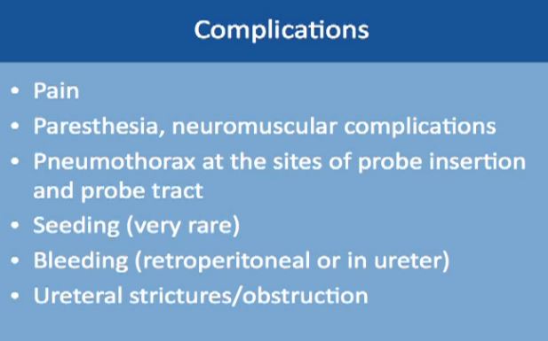
There are some complications, including pain after the procedure. This is usually an overnight stay; the patient goes home the next day. There could be some numbness or neuromuscular complications. There can be some air around the lung, if the needle had to be inserted close to the lung. This is typically monitored during the procedure.
Seeding from the tumor is exceedingly rare, maybe 1 or 2 cases, so exceedingly rare, nearly unheard of, so this should not be a concern for patients who are willing to go through this procedure. Bleeding can occur, and that‘s why we like to keep patients overnight. Also there could be some strictures or narrowing of the ureter tube that drains the kidney.
So how do we know to recommend RFA or Cryoablation?
We know RFA is considered easier, faster, and safer, but actually has a higher chance of doing damage to the collecting system, the system that drains urine from the kidney. On the other hand, CA has a slightly higher risk of bleeding compared to RF. But it is better for larger tumors, and is good for deep tumors or central tumors. You can monitor live CA with ultrasound or CT to see the ice ball actually grow and treat the whole tumor.
Partial Nephrectomy
The more invasive category is the partial nephrectomy, the actual removal of the tumor, leaving the rest of the kidney in place. The standard had been the open procedure, but nowadays, people tend to do more robotic assisted surgery. You have to have a good surgical candidate for this, both as to the patient’s anatomy and the actual mass. Less commonly used is the pure laparoscopic, just because it is technically challenging and because most hospitals have a robot available.
(VIDEO available on Youtube, about 17 minutes into clip.)
We will show a two minute clip of robotic nephrectomy surgery, courtesy of my colleague. The kidney is here, artery to the kidney here and the vein to the kidney is here. First we need to stop the blood flow to the kidney to do this procedure safely and accurately. A clamp is temporarily placed on the artery and another clamp on the vein, which will completely shut off the blood supply. Here the tumor is literally being carved out of the kidney. Here is the tumor, this is normal kidney, this is tumor, and this is the tumor over here, normal kidney here, so this is what is considered a partial nephrectomy. We check that the mass is excised and then suture the kidney so we can get normal control and prevent bleeding. All you see here is normal kidney. (This is on slightly fast forward–we don’t typically work this fast!)
So now they are bringing back the edges of the kidney together. We use some substance to prevent bleeding which will dissolve on their own: bringing the edges of the kidney back together. And you can see very early on that very little of the normal kidney is removed, just to give a margin to assure that we remove the whole tumor. We remove all the clamps to bring back normal blood supply to the kidney, so this is how the kidney looks. It is pink because it has the normal blood supply. There is no bleeding seen.
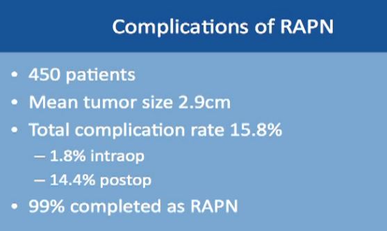
Complications might occur in some procedures. This is a large study of almost 500 robotic-assisted partial nephrectomies. Tumor sizes were about 3 cm, so small kidney masses. The total complication rate is about 16%. Complication rates include everything, even slight fevers, so higher than you would expect. Less than 2% of the complications happen during the surgery; most of the complications happen afterwards. About 99% of the surgeries were completed with a robot. Main complications specific to a robotic–assisted partial nephrectomy are bleeding, urinary leakage (1.7%), radical nephrectomy (1.6%), which means removing the whole kidney.
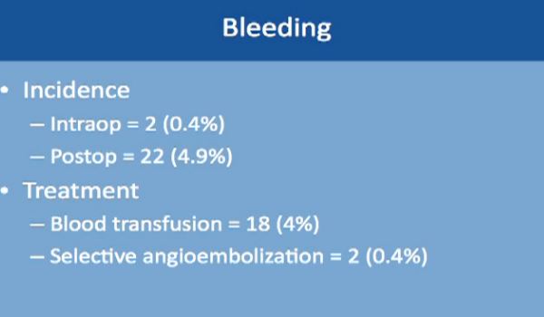
Bleeding occurred in two patients only during the operation, and about 5% of patients while they are still in the hospital. The treatment is relatively simple. Blood transfusion took care of 4% of these patients. About 2% of these patients needed selective embolization, a procedure that is usually done by interventional radiology and done under sedation. This saves the kidney and controls the bleeding as well.
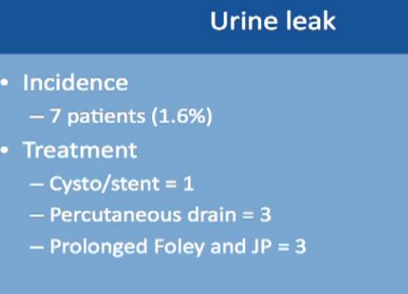
Urinary leakage is found in less than 2%, with treatment typically to put in a stent or a drain or leaving the catheter in longer. None of these patients needed repeat operations or removal of the kidney.
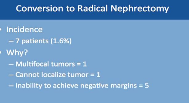
Another complication is conversion to a radical nephrectomy, which occurred in less than 1.6% of patients. This may be due to finding multiple tumors not recognized earlier. In one patient, the tumor could not be found; this happens if the tumor is small and very deep into the kidney. In five patients, a negative margin could not be achieved. That means some cancer was left behind after the tumor was carved out. Then the physician decided to remove the whole kidney to make sure all the cancer was gone. Also, one or more tumors cannot be removed successfully with the kidney left in place. Such complications occur less than 10% under any procedure.
In this table (which could not be captured), but the tumor size was about 3 cm on average. Surgery time was about 3 hours. Blood loss was about 200-300 ccs and hospital stay was about 3 days. This is a different study with complications about 5%. And with positive margins, i.e, a tiny amount of cancer was left, was about 1.6%. The follow up was about four years.
Complications after Treatment Now to compare the rate of complication with RF and cyroablations and surgery: this is from the AUA guidelines; the complication with Cryoablation is about 5%, with RFA about 6%, about 9% with lap partial, and about 6% with open partial and about 3% with complete removal laporascopically, and about 1% with open. All are less than 10% complications.
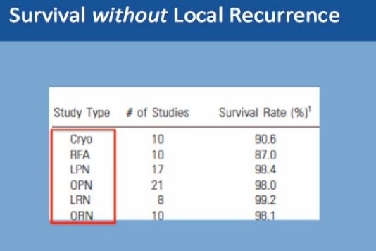
What about survival without a local recurrence?For cryoablation, it is 90% This is not complete survival, this is living without a recurrence. Again, 90% with cryolablation, with RFA about 87%, and with surgery about 98%.
Renal Function
Partial nephrectomy and RFA has similar renal functional outcomes
No change in GFR
No change in CKD stage
Kidney function is pretty much the same after these procedures, whether you do a partial removal of the tumor or an ablation, so that is not a major factor in decisions.
Active surveillance should be offered to appropriate patients. Patients should be informed that metastases could happen, but typically less than 2% of the time, and it typically takes on average 3-4 years on average. before that would happen. Half of all patients will undergo some treatment eventually, and leave active surveillance.
Survival without Local Recurrence
With the second modality, energy ablation with radio frequency or cryoablation, the chances of survival without local recurrence are about 88-90%, and we have intermediate term data, but not long term follow up on those patients.
However, with surgery you have 98% chance of survival without having a local recurrence to that same kidney. With surgery, we do have long term data as to partial nephrectomy.
A partial nephrectomy is the most definitive therapy. Any patient who is willing to undergo surgery and can tolerate surgery should do have partial nephrectomy for small renal masses. Alternatively, we can offer patients surveillance or ablation. We typically reserve that for that for those with poor kidney function—and we don’t to cause more kidney dysfunction, for those with more have multiple tumors, or for the patient at a high surgical risk or with a poor performance status. That latter patient means he might be a poor surgical candidate due to other health issues, with a heart problem or more dangerous tumors, for example. We can offer surveillance or ablation for those patients. If patient does not want surgery, we can offer two other options. This concludes my talk and I welcome questions.”
In April of 2012, the KCA held a patient conference at MD Anderson Cancer Center, which gathers about 75 patients and caregivers to hear internationally-respected experts about kidney cancer. I attended the conference and had the opportunity to meet with my “Friends on Account of Kidney Cancer”, all of joined by our experience with kidney cancer. We had never seen one another, had to send pix to make sure we could find one another, and greeted each other as long-lost friends.
To make these lectures more meaningful, I have edited each one, after countless hours of review and correction, adding the slides to the lectures and recreating them to make them more easy to email and share. Several friends through my patient connections have read them and found them useful, and I hope you will as well.
The first lecture is by Dr. Jose Karam of MD Anderson, and is presented in its edited form in the next post. Other similar posts will follow until all the lectures are available. With the ASCO conference going on, you will see mention of trials now being published presented at ASCO. These lectures will give you some skills to assess that information , which is now hitting the airways, with the usual lack of perspective and background which we need to understand these new findings.


 Those patients who had clear cell histology actually had a more favorable outcome than those with non-clear cell. Those who had advanced grade, sarcomatoid de-differentiation, those who had peri-nephrectic fat, nodal metastases, distant metastases, all were associated with a more adverse outcome than patients with venous thrombus involvement.


- A Randomized Double Blind Phase 3 Study of Adjuvant Sunitinib vs Placebor in Pts with high risk RCC")